
Specialists
Head & Neck Cancers
Cancer of the Nose and Sinuses
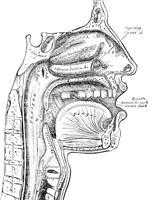
 The nasal cavity and surrounding sinuses assist in maintaining the functionality
and structure of the head and neck area, and are essential in our everyday
lives. Lined with a layer of mucous tissue, these structures are susceptible
to the abnormal cell development that can lead to cancerous tumor formation.
The nasal cavity and surrounding sinuses assist in maintaining the functionality
and structure of the head and neck area, and are essential in our everyday
lives. Lined with a layer of mucous tissue, these structures are susceptible
to the abnormal cell development that can lead to cancerous tumor formation.
Nasal cavity and paranasal sinus cancers tend to develop most often in patients who smoke, have a family history of cancer, or are frequently exposed to dust, flour, radium and other substances as part of their job. Patients with this type of cancer often experience:
- Nasal congestion
- Facial pain
- Post-nasal drip
- Nosebleeds
- Decreased sense of smell
- Watery eyes
- Vision loss
If cancer is suspected, your doctor will perform a series of tests, such as endoscopy, CT scan or biopsy to diagnose and stage your condition. Treatment for cancer of the nose or sinuses usually involves surgery to remove the tumor and surrounding tissue, which may be performed in conjunction with chemotherapy or radiation therapy to ensure complete removal. The location of each patient’s tumor is essential in determining whether or not surgery is possible.
Cancer of the Mouth and Throat Treatment
Cancer of the mouth and throat refers to the development of a cancerous tumor within the lips, cheeks, salivary glands, gums, teeth, tongue, tonsils or nearly any other area within these structures. Oral (mouth) and pharyngeal (throat) cancers most commonly affect patients over the age of 40 who smoke or chew tobacco. Excessive alcohol consumption, a family history of head and neck cancer, and exposure to the Human Papilloma Virus (HPV) may also increase a patient’s risk for mouth and throat cancer. Symptoms of oral and throat cancers can vary depending on the type and location of the cancer, but may include white patches in the mouth, a sore on the lips, bleeding, loose teeth, difficulty swallowing, earache and more.
In order to determine which type of treatment is most appropriate for each patient’s individual condition, your doctor will first determine the stage of the cancer through a series of diagnostic exams performed in our office. A customized treatment plan is then developed, which may include surgery, chemotherapy, radiation therapy or a combination of these approaches. Surgery for mouth and throat cancer involves a removal of the tumor and a small margin of surrounding tissue to ensure that all cancerous cells have been successfully eliminated.
Cancer of the Larynx Treatment
 The larynx, also known as the voice box, is located in the front of the
neck and helps us breathe, swallow and speak, as it controls the opening
and closing of the windpipe. Like nearly any other cells in the body,
abnormalities within the larynx can lead to the development of a cancerous
tumor, which most often develops within the squamous cells that line the
inner walls of the larynx.
The larynx, also known as the voice box, is located in the front of the
neck and helps us breathe, swallow and speak, as it controls the opening
and closing of the windpipe. Like nearly any other cells in the body,
abnormalities within the larynx can lead to the development of a cancerous
tumor, which most often develops within the squamous cells that line the
inner walls of the larynx.
Patients with laryngeal cancer may experience hoarseness, a lump in the
neck, a cough, difficulty breathing, earache, difficulty swallowing and
weight loss, among other symptoms. While the exact cause of laryngeal
cancer is unknown, certain people may be at an increased risk of developing
this condition, including those who:
- Are over the age of 55
- Are male
- Are African American
- Smoke
- Consume large amounts of alcohol
- Have a personal or family history of head and neck cancer
- Have been exposed to HPV
Surgical treatment for laryngeal cancer may involve endoscopic removal of tumors through the mouth using either laser or microscopic techniques, or removal of part or all of the larynx through open procedures such as a partial or total laryngectomy. If the cancer has spread from the larynx, the lymph nodes or other nearby structures may be removed as well. Radiation and chemotherapy are also commonly performed to treat laryngeal tumors, and may be used alone or in combination with surgery. A personalized, multidisciplinary treatment will be developed based on a thorough evaluation of each patient’s individual condition performed in our office.
Parotid and Submandibular Gland Tumors
 The parotid and submandibular glands make up two of the three major salivary
glands in the body that secrete saliva near the upper teeth and under
the tongue, and help aid in digestion, oral lubrication and hygiene, and
protection against tooth decay. These glands are susceptible to benign
or malignant tumors that appear as a lump in front of or below the ear.
Parotid and submandibular gland tumors can spread from other areas of
the body to enter the gland through the lymphatic system, and may include
lymphomas, melanoma or squamous cell carcinoma, among others. Fortunately,
there are several surgical procedures that can be performed to remove
these tumors.
The parotid and submandibular glands make up two of the three major salivary
glands in the body that secrete saliva near the upper teeth and under
the tongue, and help aid in digestion, oral lubrication and hygiene, and
protection against tooth decay. These glands are susceptible to benign
or malignant tumors that appear as a lump in front of or below the ear.
Parotid and submandibular gland tumors can spread from other areas of
the body to enter the gland through the lymphatic system, and may include
lymphomas, melanoma or squamous cell carcinoma, among others. Fortunately,
there are several surgical procedures that can be performed to remove
these tumors.
Parotidectomy
Treatment of parotid gland tumors may require surgical removal of the gland. This procedure, known as parotidectomy, is performed under general anesthesia and may require a short hospital stay as well. Depending on the size of the tumor, a partial or total parotidectomy may be performed. During a parotidectomy, an incision is made in front of the ear, similar to the incision used during a face lift. The tumor and a small margin of surrounding tissue are removed through the incision. After the parotidectomy procedure, a drainage tube will be placed at the incision site to collect blood, serum and saliva, which can usually be removed after one day.
Facial Nerve Monitoring
During a parotidectomy, it is important to identify the facial nerve and carefully work around it in order to reduce the risk of damage, which can result in facial paralysis or other serious complications. Facial nerve monitoring confirms the position of the nerve throughout surgery and verifies its integrity at the completion of procedure. This provides peace of mind to both patient and surgeon, ensuring that the procedure goes smoothly and reducing the chance of post-operative complications.
Submandibular Gland Excision
Surgical removal of the submandibular gland may be necessary to provide relief from tumors or chronic infection. Submandibular gland excision is performed under general anesthesia, and patients usually return home the same day. During the excision procedure, an incision is made in the neck, right under the jawbone. The entire gland is then removed from the surrounding muscles, vessels and nerves. After the submandibular gland excision procedure, a drainage tube may be placed at the site of the incision, which can then be removed the next day.

-
ENT/Otolaryngology ENT, Otolaryngology 2557 Mowry Ave.
Suite 30
Fremont, CA 94538
(510) 248-1590 More Information
View All
/
-
 Maria's Story ENT
Finding relief for a deviated septum has been a long and exhausting journey for Maria Dibs. “I fell and fractured my nose as a ...
Read Full Story -
Cal's Story ENT
“Dr Van Tassel is the best! This doctor knows what he is talking about – he not one of those doctors that play a guessing game ...
Read Full Story -
Benny's Story ENT
“This guy is great. I had been having an issue for a few years with my throat and after going through quite a few different ...
Read Full Story